
Kidney stones are notoriously painful and unpleasant. When a large kidney stone passes into the ureter, it results in an acute pain known as renal colic.
Kidney stones, or renal calculi, develop due to the abnormal formation of crystals in the urine. Kidney stones are generally categorized according to their location, with renal lithiasis referring to stones in the kidneys and ureterolithiasis referring to stones in the ureter.
Kidney stones are also categorized according to their chemical composition. They can be made of calcium, struvite, uric acid and other compounds. About 85% of kidney stones are made of calcium.
Kidney stones occur most frequently in middle-aged and older adults and are more common in men than women. The stones themselves vary in size, with the smallest being invisible to the naked eye and the largest having a diameter exceeding 2.5 centimetres.
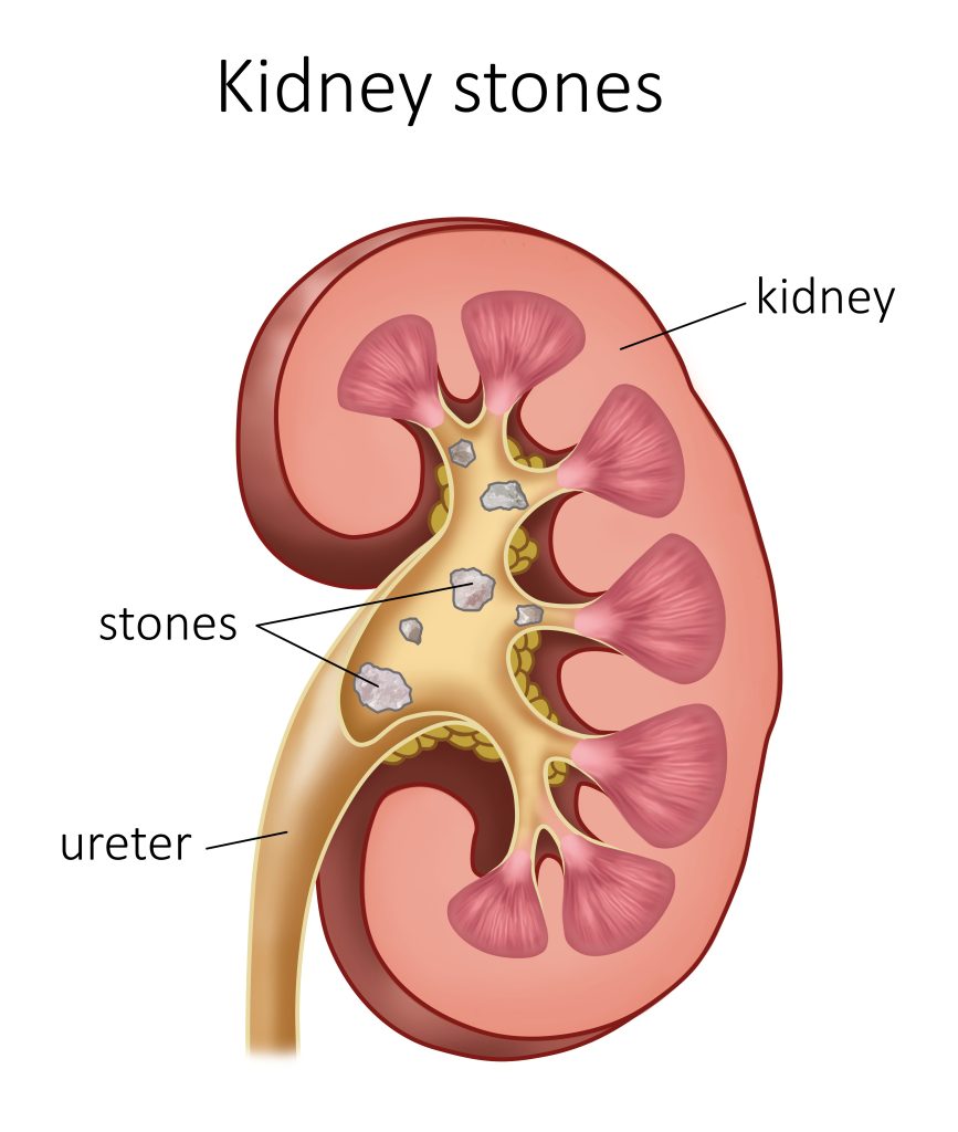
Staghorn kidney stones, which have many branches that resemble antlers, can fill nearly the entire cavity of the renal pelvis (the central funnel in the kidney) and the adjoining tubes.
What causes kidney stones?
Kidney stones are most common among individuals with hyperparathyroidism. They’re also regularly found in individuals with a diet high in animal protein or who consume too much vitamin C, too little calcium or too little water.
People with a family history of kidney stones are more likely to have kidney stones themselves.
People who’ve undergone surgery for weight loss (bariatric surgery) are also susceptible to developing kidney stones.
What symptoms of kidney stones are most common?
Kidney stones that block a ureter, the renal pelvis or one of the kidney’s drainage tubes can cause renal colic. Renal colic is characterized by intense, intermittent pain, typically in the area between the ribs and hips, that radiates into the abdomen and often the genital area.
Kidney stone pain tends to occur in waves lasting approximately 20 to 60 minutes. During each wave the pain increases to a maximum intensity then fades. The pain may travel down to the abdomen and reach the groin, testicles or vulva.
Renal colic may be accompanied by nausea and vomiting, restlessness, sweating and blood in the urine (hematuria). In addition, the affected person may feel a frequent, strong urge to urinate, in particular as the stone passes through the ureter. Expulsion of a stone or stone fragment while urinating is possible.
How are kidney stones diagnosed?
A uroscan (a type of CT scan) can be used to locate a kidney stone and determine the extent to which it’s obstructing the urinary tract.
An ultrasound is an alternative to a CT scan and doesn’t expose the patient to radiation. However, it cannot detect small stones, especially ones in the ureter.
An X-ray of the abdomen may also be taken in some cases. However, an X-ray is much less accurate in diagnosing kidney stones and can only detect calcium stones.
What treatment is available for kidney stones?
Potassium citrate may be prescribed to individuals with uric acid stones for four to six months. This mineral compound, which is taken as a pill, makes the urine more alkaline and can gradually dissolve uric acid stones.
Small stones that don’t cause urinary tract obstruction, infection or other symptoms generally don’t require treatment and are ultimately expelled without intervention.
Larger stones (greater than five millimetres in diameter) and stones located near the kidney are less likely to be expelled spontaneously.
Certain medications such as Tamsulosin and calcium channel blockers may increase the chances of spontaneous stone expulsion.
Shock wave lithotripsy can be used to break up a stone in the pelvis or upper ureter that’s no more than one centimetre in diameter. In this procedure, low energy acoustic wave pulsations are directed at the stone to fragment it. The stone fragments are then eliminated in the urine.
A semi-rigid ureteroscopy can be used for kidney stones located in the lower segment of the ureter.
A flexible ureteroscopy can be used for kidney stones located in the upper ureter and renal pelvis.
Another treatment option is a percutaneous nephrolithotomy. This surgical procedure involves making a small incision in the patient’s back and inserting an optical tube called a nephroscope into the kidney. Once the tube is in place, the doctor inserts a probe through it to break the stone into smaller fragments and remove them.
What treatments are available at Cliniques Marois?
- Semi-rigid ureteroscopy treatment
- Flexible ureteroscopy treatment