
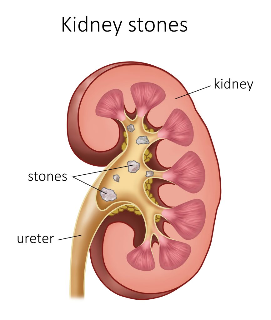
Ureteroscopy is needed to remove a stone/stones from the kidneys, which have not been able to normally pass through the ureter (connection between the kidney and the bladder).
Ureteroscopy involves the passage of a small fibroptic instrument called a ureteroscope through the urethra and the bladder into the ureter. This 3 mm diameter optical instrument is connected to a light source, sterile water irrigation and a camera. It also contains a working channel through which various instruments are introduced, such as metal guides, laser fibers and basket probes. As such, Dr Marois can actually look into the ureter, find the stone and remove it.
The manipulation of the instruments is done under direct vision and by fluoroscopic guidance (by X-rays).
")
Why do a flexible ureteroscopy for a kidney stone?
Ureteroscopy can be a second attempt to intervene if this first alternative fails. It can also serve in a cancer diagnosis or in the treatment of ureteral abnormalities such as stenosis (narrowing of channels or vessels).
The purpose of the intervention is to fragment the stone with a laser fiber. The fragments are left in place or can be removed by a tiny basket probe.
The treatment is usually very effective.
How is a flexible ureteroscopy performed for a high renal stone?
The procedure is performed under spinal anesthesia at the Centre Métropolitain de Chirurgie. The patient is placed in a lithotomy or gynecological position.
It requires the use of a radiology device to perform fluoroscopy at the same time to, among other things, orient and assess the fragmentation by laser (C-ARM). It is handled by a radiology technician at the same time.
A rigid cystoscope is introduced into the ureter under direct vision down to the level of the bladder. The ureteral opening is identified and cannulated by a catheter to introduce a first safety metal guide to the level of the renal cavities and pelvis. A second work guide is introduced using a double lumen catheter.
An ureteral access sheath is usually introduced until the stone. The ureteroscope is then easily inserted inside this catheter and then a laser fiber is introduced into the working channel of the ureteroscope. The kidney stone is fragmented progressively under direct vision. Controlled pressure water irrigation is necessary to ensure proper visibility. If visibility is compromised by bleeding, the surgery is stopped. It is also possible that the fragmentation of the calculation is incomplete. A second session or other alternatives may be offered in the following weeks.
At the end of the procedure, it is generally necessary to leave in place a double J in order to avoid lower back pain of the renal colic type, secondary to the obstruction caused by the edema, which is the latter with no relation to the calculation. The double J can be left in place a few days to a few weeks after the procedure.
What are the risks and possible complications of a flexible ureteroscopy?
The most common difficulty (10%) is the failure of progression in the ureter, caused by too narrow ureteral size. This is why the need for several operating times is always considered in cases where the ureter is not prepared beforehand by a double catheter J or if the computation to be treated is bulky. To avoid the risk of trauma, a double J is temporarily installed so that the ureter expands on it. It is then necessary to postpone the intervention and to re-intervene a few weeks later.
The discomforts secondary to double J are common. An infection or sepsis is possible despite sterile urine and preoperative antibiotic prophylaxis. They can occur quickly the same day or late.